3D reconstruction for diagnostics has revolutionized radiology and surgery by enabling detailed visualization of internal structures, enhancing accuracy in detecting abnormalities, improving communication among healthcare professionals, facilitating early detection and diagnosis, and ultimately leading to better patient outcomes through more precise planning and minimal invasive procedures.
“The integration of 3D imaging technologies into radiology and surgery has revolutionized healthcare, significantly enhancing diagnostic accuracy and patient outcomes. This article explores the multifaceted applications of 3D reconstruction in medical practices. From enhancing diagnostic capabilities through detailed three-dimensional views to aiding surgeons in navigating complex anatomies, 3D imaging is transforming care. We delve into how this technology visualizes internal structures, enables comprehensive patient management, and even influences interventions, setting new standards in modern medicine.”
Enhancing Diagnostic Accuracy with 3D Reconstruction
In radiology, 3D imaging has transformed diagnostic capabilities by enabling detailed visualization of internal structures. Through advanced 3D reconstruction techniques, radiologists can create accurate, multidimensional representations of organs and tissues, enhancing their ability to detect abnormalities that might be obscured in traditional 2D images. This technology is particularly valuable in complex cases where understanding the precise anatomy is crucial for effective treatment planning.
The process involves reconstructing a series of 2D images into a unified 3D model, allowing for comprehensive assessment from multiple angles. This not only improves diagnostic accuracy but also facilitates better communication between healthcare professionals. By providing a more complete picture, 3D reconstruction aids in the early detection and diagnosis of various conditions, ultimately leading to improved patient outcomes.
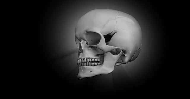
Navigating Complex Anatomy: 3D Imaging in Surgery
Navigating complex anatomy has always been a significant challenge in surgery, particularly with intricate structures and limited visual access. Traditional 2D imaging often falls short in providing a comprehensive understanding of the three-dimensional (3D) nature of organs and tissues. However, 3D reconstruction for diagnostics has emerged as a game-changer in this domain. By utilizing advanced imaging techniques like computed tomography (CT) and magnetic resonance imaging (MRI), healthcare professionals can now create detailed 3D models of anatomical regions of interest.
These models offer unparalleled visualization capabilities, allowing surgeons to explore every nook and cranny of the body from multiple perspectives. This level of detail enables more precise planning for complex surgical procedures, improves decision-making during operations, and ultimately enhances patient outcomes. Moreover, 3D imaging aids in identifying subtle abnormalities or structures that might be obscured by traditional 2D views, fostering a deeper understanding of the anatomy and leading to more effective interventions.
Visualizing Internal Structures for Comprehensive Care
In radiology, 3D imaging has transformed the way internal structures are visualized and understood. Through advanced techniques like 3D reconstruction for diagnostics, healthcare professionals can gain a more comprehensive view of the human body. This enables them to make more accurate diagnoses and plan treatments with greater precision. By rendering complex anatomical data into three-dimensional models, radiologists can identify subtle abnormalities that might be overlooked in traditional two-dimensional imaging.
For surgery, these technologies offer equally profound benefits. Surgeons can use 3D reconstructions to map out critical structures, such as blood vessels and nerve pathways, before operating. This preoperative planning allows for more informed decision-making during procedures, minimizing the risk of complications and enhancing overall patient care. The detailed visualization enabled by 3D imaging facilitates safer surgeries and contributes to better clinical outcomes.
Revolutionizing Interventions with Advanced 3D Technology
In the realm of radiology and surgery, advanced 3D technology is revolutionizing interventions with its unprecedented level of detail and accuracy. 3D reconstruction for diagnostics allows healthcare professionals to gain a comprehensive view of internal structures, enabling more precise planning and execution of medical procedures. This innovative approach goes beyond traditional 2D imaging by providing a detailed, three-dimensional model of the patient’s anatomy.
Such technology offers numerous advantages in both diagnostic and surgical settings. For instance, it enables radiologists to detect subtle abnormalities that might be overlooked on standard X-rays or CT scans. Similarly, surgeons can better navigate complex procedures with visual guidance, ensuring minimal invasiveness and enhanced outcomes for patients.
3D imaging has revolutionized both radiology and surgery, offering unprecedented accuracy and insights. By leveraging 3D reconstruction for diagnostics, healthcare professionals can navigate complex anatomy with greater ease, visualize internal structures more comprehensively, and ultimately enhance patient outcomes. Advanced 3D technology continues to push the boundaries of what’s possible, ensuring a future where interventions are more precise and effective.